What It Does
Gestalt Mitotic Figure Detection supports pathologists by identifying and highlighting mitotic figures across routine H&E whole-slide images from various carcinomas, sarcomas, and cutaneous tumors.
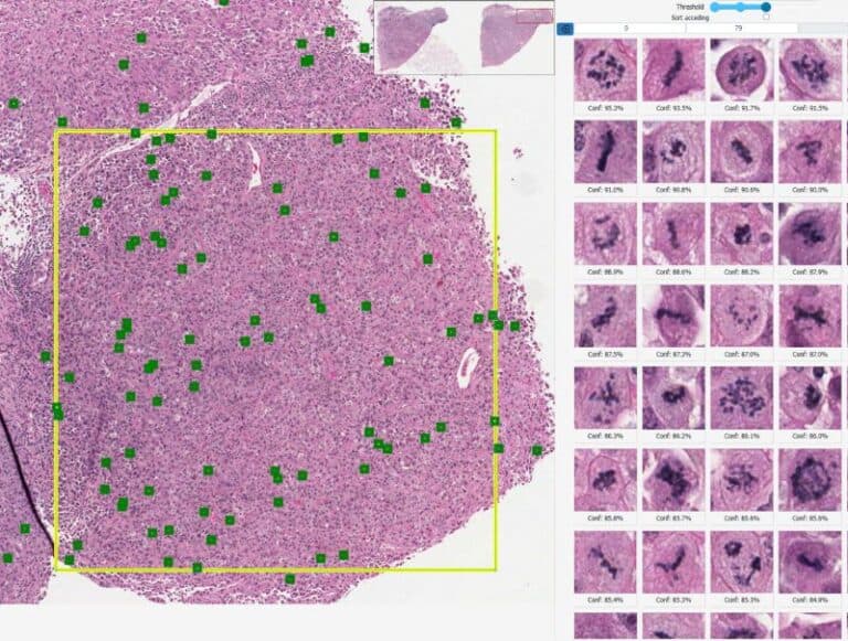
The algorithm analyzes the entire slide at high resolution and automatically calculates the 10 HPF region with the highest mitotic figure activity. This surfaces the most diagnostically relevant candidates to assist with rapid review, improved consistency, and reduced manual workload during routine assessment.
How It Works
Analyzes routine H&E-stained whole-slide images
Scans the entire slide at high resolution
Identifies the 10 HPF area with peak mitotic density
Detects and highlights candidate mitotic figures
Presents results directly within the digital workflow for pathologist review